Digital Infrared Thermography
Digital Infrared Thermography The Only Method Available for “Visualizing” Your Pain.
An FDA approved device.
What is Infrared Imaging?
Digital infrared thermal imaging is a totally non-invasive clinical imaging procedure for detecting and monitoring several diseases and physical injuries, by showing the thermal abnormalities present in the body. It is used as an aid for diagnosis and prognosis, as well as monitoring therapy progress, for conditions and injuries, including:
-
Back Injuries
-
Arthritis
-
Headache
-
Nerve Damage
-
Unexplained Pain
-
Fibromyalgia
-
Breast Disease
-
Carpal Tunnel Syndrome
-
Disc Disease
-
Inflammatory Pain
-
Skin Cancer
-
Referred Pain Syndrome

Images or ‘Thermograms’ can be taken of the whole body or just areas being investigated. The digitized images are stored on a computer and are sent electronically to a Thermologist (certified doctor) for interpretation and reporting. Your report is color printed and a copy can also be sent to your healthcare professional.
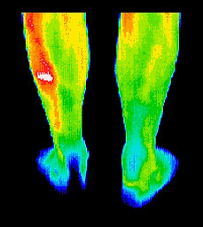
Football player with a stress fracture that was not detected with X-Ray.
The bone scan confirmed DITI findings.

Upper back and neck pain, also regular headaches. Thermography guided treatments achieved better results than any previously.

Pain in the jaw. Thermogram findings helped confirm the diagnosis of TMJ and referral to the appropriate specialist for treatment.

Significant inflammatory changes in the right breast. Referral to a breast specialist and subsequent biopsy diagnosed inflammatory breast cancer at a very early stage

Arthritic disorders generally appear as “hot areas”, since the affected sites are usually inflamed.

A painful ankle injury can be monitored throughout treatment and rehabilitation.
Procedure
Unlike most diagnostic tests D.I.T.I. is:
-
Noninvasive
-
No radiation
-
Painless
-
No contact with the body
F.D.A approved
-
This quick and easy test starts with your medical history being taken before you partially disrobe for the scanning to be performed. Standard region of interest exams approximately 15 minutes and a full body 30 minutes. Your printed report is normally completed within a few days.
CLINICAL D.I.T.I. is providing the answers in the diagnosis of pain.
-
The only method available for visualizing pain and pathology.
-
Can assess pain and pathology anywhere on the body.
-
Is a very useful adjunctive procedure to other diagnostic tools.
-
Is very cost-effective, risk-free, and provides instant images.